crystal violet biofilm
The cultivation of the strains should be conducted in a similar way like the crystal violet biofilm assay but without long-term cultivation for biofilm formation. However 96 well microtitre plate based assays share the issue of edge effect.

All Positive Cultures Demonstrated Staining For Crystal Violet Download Scientific Diagram
Wash 4X with 3ml H2O gently to remove unbound stain 6.

. The crystal violet visualized the biofilm biomass reduction of 94 60 and 67 for 24 h 48 h and 72 h biofilm respectively when a. Two assays for quantification of S. In CV assay most isolates formed weak biofilm 743 while the rest formed moderate biofilm 233 or strong biofilm 23.
Crystal violet CV staining a colorimetric technique has been utilized broadly to measure biofilm development in part due to its amenability to huge screening methods 25 26. In doing so high moderate and non-. Using partial least square.
First 01 crystal violet is prepared and the biofilm-formed specimen is immersed in it for 30 min after which the specimens surface is rinsed with tap water. 15 hours agoBiofilm formation in ocular bacteria and fungi by the tissue culture plate method using crystal violet method. Row B of 58 wells 4 replicates of MRSA-3S strain.
Then 200 μl of crystal violet solution 02 was added to all wells. Add 125 μL of a 01 solution of crystal violet in water to each well of the microtiter plate. Row B of 14 wells 4 replicates of MRSA-44S strain.
The objective of this study was to optimize the Crystal Violet phenotypic biofilm screening technique for S. Raman spectroscopic analysis of the bacteria revealed most spectral differences between high and low biofilm performers in the fingerprint region between 750 and 1150 cm 1. As edge effect causes a significant increase in plate.
Fixed with 99 methanol. The colors selected for the epifluorescence microscopy data reflect the. The primary cause of the edge effect phenomenon is evaporation.
Remove media from biofilms and wash 1X in 1ml PBS 2. Biofilms were stained with crystal violet dissolved in 200 μL of 10 acetic acid. The time course of biofilm growth must be determined empirically for each organism and set of conditions used.
Crystal Violet Protocol for Biofilms 1. 9 who reported the maximal OD 600 mean values of approximately 2 following crystal violet. Biofilms are communities of microbes attached to surfaces which can be found in medical industrial and natural settings.
Bivia A or a multi-species biofilm composed of all three species B using the crystal violet method total cell counts by epifluorescence microscopy and the colony-forming units CFU method. After the formation of the biofilm on microtiter plates the supernatant of the wells was discarded. Therefore the bacterial strains were grown in TSB at 160 rpm and 37C using an overnight culture for inoculation with an OD of 005.
Comparison of biofilm quantification by 05 crystal violet Grams crystal violet and safranin. Figure 1 Quantification of 24 h and 48 h single-species biofilms of G. However most isolates in.
Early phase biofilms are also prone to damage by the latter steps. After aspiration of planktonic cells biofilms were. For most applications particularly verification assays for surface area adhesion-deficient mutants 25 27 calculating the absolute quantity of biofilm shaped by.
The value of each biological replicate is the mean of. Crystal violet staining assay of methicillin-resistant Staphylococcus aureus strains having different biofilm formation capability. Microplates are essential tools for biofilm research since it allows high throughput screening of biofilm forming strains or in the assay of anti-biofilm drugs.
Add 1ml 04 Crystal Violet stain to each biofilm and let sit room temp 45min 4. Biofilms were grown in microtiter plates in tryptic soy broth TSB or brain heart infusion BHI at 30 C for 24 or 48 h and quantified via the crystal violet assay. In fact life in a biofilm probably represents the predominate mode of growth for microbes in most environments.
Remove Crystal Violet stain 5. Crystal violet CV assay is the most popular method for biofilm determination adopted by different laboratories so far. SE1457 strain was exposed to various concentration of nicotine 0 0025 005 05 5 50 500 μgml for 48 h.
The eventual decrease in crystal violet staining is presumed to occur because the lack of nutrients may stimulate the bacteria to detach from the surface Sawyer and Hermanowicz 1998. Each symbol shows the mean of 4 biological replicates prepared from individually grown cultures. Each strain was evaluated in triplicate.
Aureus DSM20231 and S. However biofilm layer formed at the liquid-air interphase known as pellicle is extremely sensitive to its washing and staining steps. Evaluation of the Biofilm Formation with Crystal Violet Assay.
A significantly larger of. Up to 10 cash back Two assays for quantification of S. Let biofilms air dry 45min room temp 3.
32 Quantification of Biofilm Mass by Crystal Violet Staining Assay. In this study the quantity of biofilm as shown by crystal violet staining increased in a time-dependent manner to reach maximal OD 600 mean values of approximately 12 on the 7th and 8th day. Plates are washed twice with phosphate buffer saline or sterile saline.
Row A of 58 wells 4 replicates of MRSA-51S strain. Medium was removed from the wells which were washed three. The color change in the materials surface is evaluated by using a color meter CR-13 Konika-Minolta Sensing Co.
Aureus biofilm formation the crystal violet CV assay and the XTT tetrazolium salt reduction assay were optimized evaluated and further compared. In CV assay most isolates formed weak biofilm 743 while the rest formed moderate biofilm 233 or strong biofilm 23. Aureus biofilm formation the crystal violet CV assay and the XTT tetrazolium salt reduction assay were optimized evaluated and further compared.
Included were low OD 10 medium OD 10 and 20 and high OD 20 biofilm performers as determined by the crystal violet test. Sterile 96-well polystyrene plates were inoculated with 200 μL of an initial bacterial suspension 10 5 CFUml in BHI medium and incubated at 37C for 24 and 48 h without shaking. Violet crystal staining assay allows for the measurement of a biofilm s total cell biomass comprised by the extracellular matrix living cells and dead cells.
Add 2ml 100 EtOH to each biofilm and let sit. Row A of 14 wells 4 replicates of MRSA-12S strain. Hunt et al 2004.
Biofilm formation was monitored in ocular isolates of S. This value is in contrast to the study by Ristow et al. 5 min the excess crystal violet was removed and plates were washed twice and.
Aureus L-1054-20192 S. Biofilm response of an isolate12 Through this method an isolate can be classified as high moderate or non-biofilm producer. Ltd Tokyo Japan and their L a and b values were used for.

Part Bba K1510214 Experience Parts Igem Org
Pdf An Improved Crystal Violet Assay For Biofilm Quantification In 96 Well Micro Titre Plate

Antibiofilm Formation Assay A The Graph Shows The Percentage Download Scientific Diagram
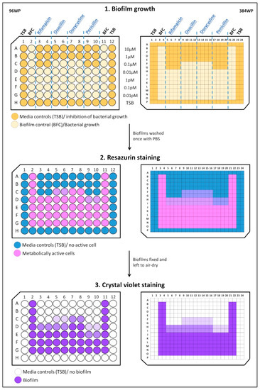
Ijms Free Full Text Optimization Of A High Throughput 384 Well Plate Based Screening Platform With Staphylococcus Aureus Atcc 25923 And Pseudomonas Aeruginosa Atcc 15442 Biofilms Html
2

Absorbance Values Of Crystal Violet Solutions Obtained From C Albicans Download Scientific Diagram

Assessment Of Biofilm Biomass By Crystal Violet Staining And Cell Download Scientific Diagram

Figure 1 Screening Of The Extent Of Biofilm Production By Tissue Culture Plate Method Tcp High Moderate And Non Slime Producers Differentiated With Crystal Violet Staining In 96 Well Tissue Culture Plate

Figure 3 From Quantitative And Qualitative Assessment Methods For Biofilm Growth A Mini Review Semantic Scholar
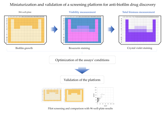
Ijms Free Full Text Optimization Of A High Throughput 384 Well Plate Based Screening Platform With Staphylococcus Aureus Atcc 25923 And Pseudomonas Aeruginosa Atcc 15442 Biofilms Html
2
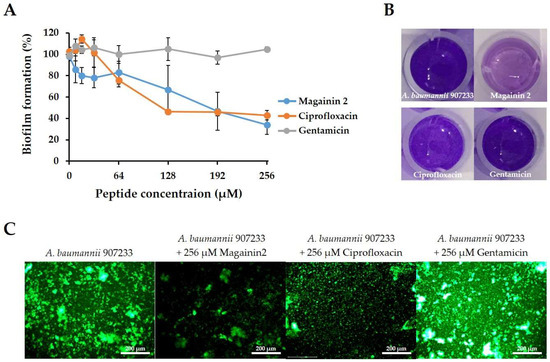
Ijms Free Full Text Antibacterial And Antibiofilm Activity And Mode Of Action Of Magainin 2 Against Drug Resistant Acinetobacter Baumannii Html

Crystal Violet Is A Stain Picked Up By The Biofilm A Crystal Download Scientific Diagram

Types Of Biofilms Formed By S Typhimurium Sv5015 Grown In Different Download Scientific Diagram

Crystal Violet Assay To Assess The Antibiofilm Activity Of Samples Download Scientific Diagram
2

Part Bba K1404008 Parts Igem Org
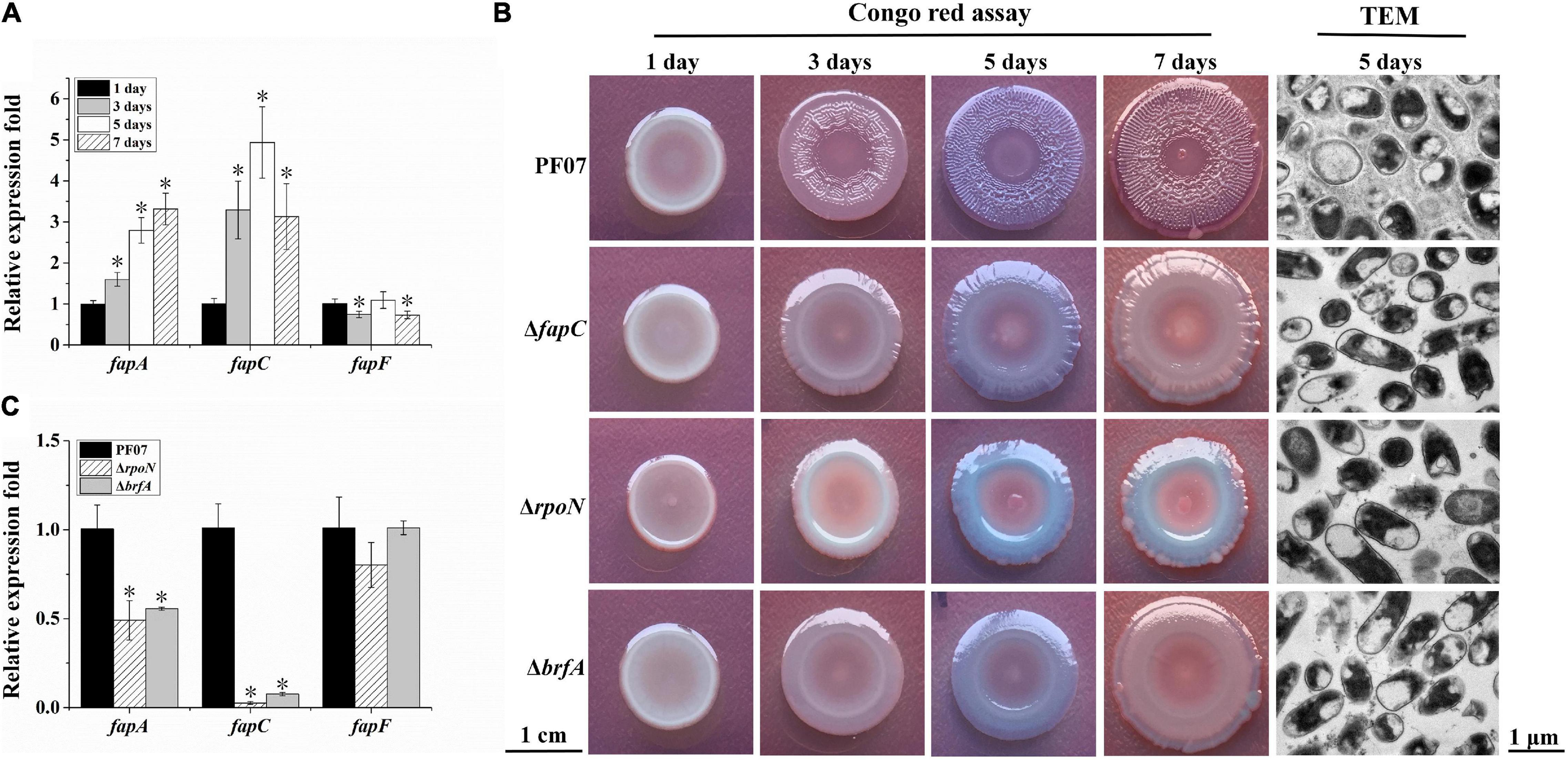
Frontiers Genes Involved In Biofilm Matrix Formation Of The Food Spoiler Pseudomonas Fluorescens Pf07 Microbiology

Quantification Of Biofilm Formation By The Pao1 Dpa3731 And Dpa3731comp Strains Using A The Crystal Violet Test B The Biofilm Ring Test Control Without Bacteria C Adherence To Human A549 Pneumocyte Cells


Comments
Post a Comment